

Introduction
Microneedles and their types
Disease detection
Detection of signaling molecules
Detecting metabolites
Monitoring physiological conditions
Conclusion and future prospective
Introduction
Agriculture is crucial to the global economy, and worries regarding its sustainability are becoming more recognized (Lew et al., 2020). In the current scenario, farmers are facing numerous challenges, including severe heat, soil degradation, drought, and plant diseases which are expected to worsen with climate change (Roper, et al., 2021). To address these challenges, plant health monitoring offers an effective approach to increasing yields and reducing environmental impact. By utilizing low-cost in-field methods, farmers can regularly monitor water levels, soil quality, and the presence of pathogens and pests. This approach allows for rapid interventions, optimizing resource use, and boosting crop output while minimizing environmental impact (Martinelli et al., 2015). The advancement of sensor technologies for early diagnosis is vital since it allows the detection of hazards (Mulimani et al., 2024). Current diagnostic technologies such as deoxyribonucleic acid (DNA) amplification methods using polymerase chain reactions (PCR) (Negahban et al., 2024), loop-mediated isothermal amplification (LAMP) (Wanjala et al., 2024), enzyme-linked immunosorbent assays (ELISA) (Nazir et al., 2024), immunofluorescence (Baysal-Gurel et al., 2008), fluorescence in situ hybridization (FISH) (Milner et al., 2019), and flow cytometry (FCM) (Jyoti et al., 2024) are limited to the laboratory settings (Mane et al., 2023). They require skilled personnel for operation, lack continuous monitoring capabilities, and are often inaccessible in remote locations (Martinelli et al., 2015). Therefore, developing compact, fast, cost-effective, and portable devices for plant health monitoring is crucial. These devices should provide initial insights into crop health without the delays associated with laboratory sample extraction and testing. Achieving sensitive, rapid, and early diagnosis of plant health is essential for enhancing productivity, protecting crops, and ensuring sustainability, food security, and competitiveness in agriculture.
Recently, microneedle (MN) technology has seen widespread use in agriculture (Wang et al. 2013). Integrating MN technology with smart sensing systems enables real-time monitoring of plants and the agricultural environment, providing valuable insights for improved agricultural practices. Smart MN based sensing systems are revolutionizing sustainable agriculture with their minimally invasive and highly sensitive capabilities. Compared to traditional sensing platforms, MN sensor systems enable immediate analysis of abiotic and biotic threats, thereby enhancing plant health. Due to their minimal impact, they can be used repeatedly on the same plant, allowing for long-term monitoring without causing significant stress. They can be integrated into continuous monitoring systems, providing rapid responses without the need for manual sampling and testing. MN sensors excel at detecting very low concentrations of analytes thanks to their high surface area-to-volume ratio. This makes them highly sensitive and capable of identifying trace amounts of nutrients, pathogens, or stress markers in plants. They can also be integrated with Internet of Things (IoT) platforms for remote monitoring and data analysis, significantly improving the management of large-scale agricultural operations. The detailed data provided by these sensors can be customized for different crops and environmental conditions, enabling precise applications of water, fertilizers, and pesticides. This optimization reduces resource waste and enhances efficiency. Furthermore, MN sensors can monitor specific plant parts, such as leaves and stems, or particular soil regions, providing localized data for more effective interventions. By facilitating precise and timely interventions, microneedle sensing technology helps reduce the overuse of chemicals and water, thereby promoting more sustainable agricultural practices. Typically, the MN platform consists of methodically ordered micron-sized arrays on a tiny patch. These MN arrays can be fabricated in various geometries, such as pyramid and conical shapes, with lengths ranging from 25 to 2000 µm and widths from 50 to 250 µm. MNs are fabricated using diverse methods including 3D printing (Parrilla et al., 2024), dry and wet etching processes (Eş et al., 2024), micro milling (Kundu et al., 2019), and laser ablation (Shahriari et al., 2024). MN technology has been combined with nucleic acid amplification platforms for the swift recognition of plant illnesses and inspection of plant pathogens. Furthermore, MNs are utilized in agriculture for the ongoing monitoring of critical physiological plant features. This includes the detection of polyphenolic levels (Dhanjai et al., 2020), water transport status (Baek et al., 2018), abscisic acid (Wang et al., 2021) and salicylic acid levels, and pH monitoring (Hossain and Tabassum, 2022).
The application of MNs in biosensing is an emerging field with limited literature available in agriculture. There is a pressing need for more consistent MN based detection approaches to offer early responses to farmers and food control organizations. To develop more accurate detection tools for agriculture, further investigation into the design parameters of MNs is essential. Thus, the current review highlights the significance of MNs as a capable alternative and integrated tool in agrotechnology, offering a new assessment in plant health monitoring. The review covers various aspects of MN technology including design, geometry, materials, and fabrication techniques of MN arrays. It examines current research trends and discusses challenges associated with applying MN technology in agricultural biosensing.
Microneedles and their types
The idea of MNs was first proposed in 1976 (Ece et al., 2023). However, it wasn’t until late 1998 that MNs were experimentally introduced as a substitution for transdermal drug delivery (Henry et al., 1998), largely due to constraints in microfabrication techniques. Early research primarily focused on animal models, but recent research advancements in MN fabrication have accelerated their adoption across various sectors, particularly in agriculture. Unlike conventional methods, MNs’ minimal invasive nature reduces the impact on plant health during insertion and allows for their reuse in on-site applications, making them advantageous for farms (Viswan et al., 2022). The agriculture sector has recently embraced MNs for treating plant illnesses, monitoring plant health, and detecting pathogens and agrochemicals. Traditional tools for treatment, inhibition, and biosensing in agriculture are often invasive and inefficient, relying heavily on agrochemicals or ineffective biomarker capture methods (Zhang et al., 2024). To enhance effectiveness, precision, and speed, MNs have undergone iterative modifications in their type, material, and geometry. Key architectural characteristics of MNs such as height, aspect ratio, base diameter, tip angle, and radius of curvature are pivotal in their design, influencing how MNs interact with target tissues (Makvandi et al., 2022). For example, MNs with a greater aspect ratio can better penetrate the plant cuticle, reaching deeper layers (Ece et al., 2023). The shape of MNs likewise affects their penetration capability, shorter MNs may not penetrate as deeply, while longer MNs can reach deeper layers (Manoj et al., 2020). Additionally, the spacing between MN arrays is critical; wider spacing may impede sufficient fluid sampling from plants, complicating the accurate detection of target analytes. Researchers are optimizing these features through various fabrication techniques to increase penetration in certain plant regions. The choice of fabrication method, material, and design parameters significantly impacts the efficacy of treatment and detection processes.
MNs are generally classified into four categories: solid, coated, hollow, and dissolving MNs (Fig. 1). The design of MNs depends on the anticipated geometric features, achieving higher aspect ratios and lower tip angles requiring more advanced techniques and materials. The constituents of solid MNs are critical as they influence their mechanical strength, reusability, and biocompatibility. Silicon is a popular choice for solid MN construction due to its excellent biocompatibility and mechanical strength (Wei-Ze et al., 2010). Alongside silicon, metals such as tungsten (W) (Ma et al., 2016), titanium (Ti) (Li et al., 2017), and stainless steel (Dhanjai et al., 2020), also polymers like poly(methyl methacrylate) (PMMA) (Ju et al., 2020) and poly (methyl vinyl ether) (PMVE) (D’Amico et al., 2023), are widely used. Biocompatible materials such as chitosan (Gorantla et al., 2021) and poly(lactic acid) (PLA) (Cha et al., 2014) are also well-suited for solid MNs. Different microfabrication methods are adapted to process specific materials. Microelectromechanical systems (MEMS) are capable of manufacturing solid MNs with the most favorable design parameters for agricultural applications (Zhang et al., 2009). However, this process is complex, requiring cleanroom processes involving thin film deposition, photolithography, and etching. Although precise, MEMS manufacturing is labor-intensive, time-consuming, and costly. Hence, to overcome these challenges, cleanroom-free fabrication approaches have been explored including microinjection molding (Gao et al., 2017), CO2 laser cutting (Chen et al., 2020), hot embossing (Li, Zhou, et al., 2019), drawing lithography (Chen et al., 2018), electroplating (Miller et al., 2015), micromilling, and spray deposition (Kim et al., 2018), 3D printing techniques (Krieger et al., 2019), and two-photon polymerization (Rad et al., 2021) have been exploited. These methods simplify the manufacturing methods while maintaining their precision and adaptability to various materials and designs, enabling the production of MNs for diverse agricultural applications.
Coated MNs are produced by immobilizing coating agents on their tips (Tarbox et al., 2018). The size and geometry of the MN arrays are crucial, as these factors determine the amount of coating agent that can be immobilized on the surface. When coated MNs interact with the plant’s interstitial fluids, the coating agent captures electrochemical or chemical signals to monitor physiological alterations. The fabrication methods for coated MNs are similar to those used for solid MNs but include an additional immobilization step. Various coating processes have been reported, with the most common methods being inkjet coating (O’Mahony et al., 2017), dip-coating (Liang et al., 2020), and spray-coating (Ning et al., 2020). This process ensures that MNs are effectively coated, enabling them to function efficiently in detection.
Advancements in microfabrication techniques have made the manufacturing of hollow MNs increasingly prevalent. Hollow MNs are particularly useful in detection studies as they can collect necessary samples from plants (Parrilla et al. 2024). Materials such as silicon (Li et al., 2019), metals (Dong et al., 2023), polymers (Dardano et al., 2021), and glass (Hu et al., 2020) are widely used in constructing hollow MNs. Traditionally, the MEMS technique, followed by deep reactive ion etching (DRIE), is employed for mass producing MNs (Ashraf et al., 2010). In addition, various advanced fabrication methods have been employed including 3D laser lithography (Suzuki et al., 2018), CNC machining (Badnikar et al., 2022), micro-stereolithography (Kim et al., 2016), and UV lithographic processes (Wang et al., 2013). These hollow MNs can extract a larger sample volume than other types of MNs, significantly improving the detection limits and analysis accuracy.
Dissolving MNs have gained significant notice in recent years due to their superior properties, such as simple manufacturing and easy application (Sartawi et al., 2022). These MNs are primarily used for drug delivery applications rather than biosensing. The fabrication process of dissolving MNs typically includes a two step approach. Initially, a solid MN is produced using the abovementioned microfabrication techniques, serving as a male template. Then, polydimethylsiloxane (PDMS), a commonly used polymer, is poured over a male template to form female PDMS molds. These molds are cured on a hot plate at ~80°C for about 2 h. Lastly, a polymer solution is transferred into the cured female PDMS mold and polymerized under specific conditions, which include monomer concentration, temperature, cross-linking agents, polymerization time, and solvent (Badnikar et al., 2020). These dissolving MNs are classified as polymeric MNs and have attracted significant interest in polymer research. The most frequently used materials include water-soluble smart materials and biodegradable polymers, such as polyvinyl alcohol (PVA) and gelatin (Meng et al., 2020). Additionally, Table 1 describes the MN fabrication methods based on the materials used.
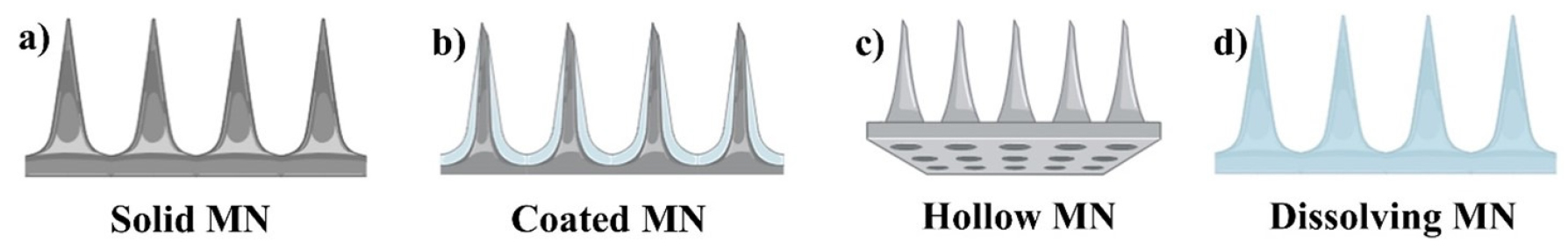
Table 1.
Information of fabrication methods of MN based on the type of material used.
Disease detection
Late blight disease, caused by the Oomycete plant pathogen Phytophthora infestants, poses a significant threat due to its rapid spread under favorable conditions, potentially devastating crops within days (Leesutthiphonchai et al., 2018). Current detection techniques involving CTAB extraction methods are limited by their need for time-consuming amplification tests and cumbersome sample preparation steps (Li et al., 2023). Recently, MNs have been employed either directly as biosensors or for on-site extraction of chemical components for further downstream analysis (Ece et al., 2023). For instance, nucleic acids have proven as key targets for diagnosing various plant illnesses and can be effectively identified using MNs (Paul et al., 2022). With this concept, MN patches made from PVA using a laser ablation strategy are explored to detect P. infestants in tomato plants (Paul et al., 2019). These MNs feature a conical cavity structure with a height of 800 µm, a base radius of 150 µm, and tip radius of 5 µm (Fig. 2(a) and (b)). Furthermore, MN patches are gently applied to the surface of the tomato leaves with a piercing force, allowing the needles to penetrate plant tissues in a minimally intrusive manner. This penetration shatters hard-to-lyse plant cell walls, and as the needle enters plant tissue, it absorbs and accumulates DNA and other molecules on the tip. Upon retraction, this procedure allows for DNA extraction in 1 min, which is faster, simpler, and less damaging (Fig. 2(c) and (d)) than CTAB methods, which typically require 3-4 h and a complex extraction protocol. The isolated DNA was then subjected to nanodrop measurement and PCR analysis, yielding a 100% detection rate. While the study demonstrates the potential of MN patches for in-field molecular diagnostics, further research is needed to evaluate their usability across a wide range of field conditions, plant species, and real-world farming environments to validate their practical application. Additionally, the current DNA extraction method uses a one-use polymeric MN patch, which may limit scalability and cost-effectiveness.
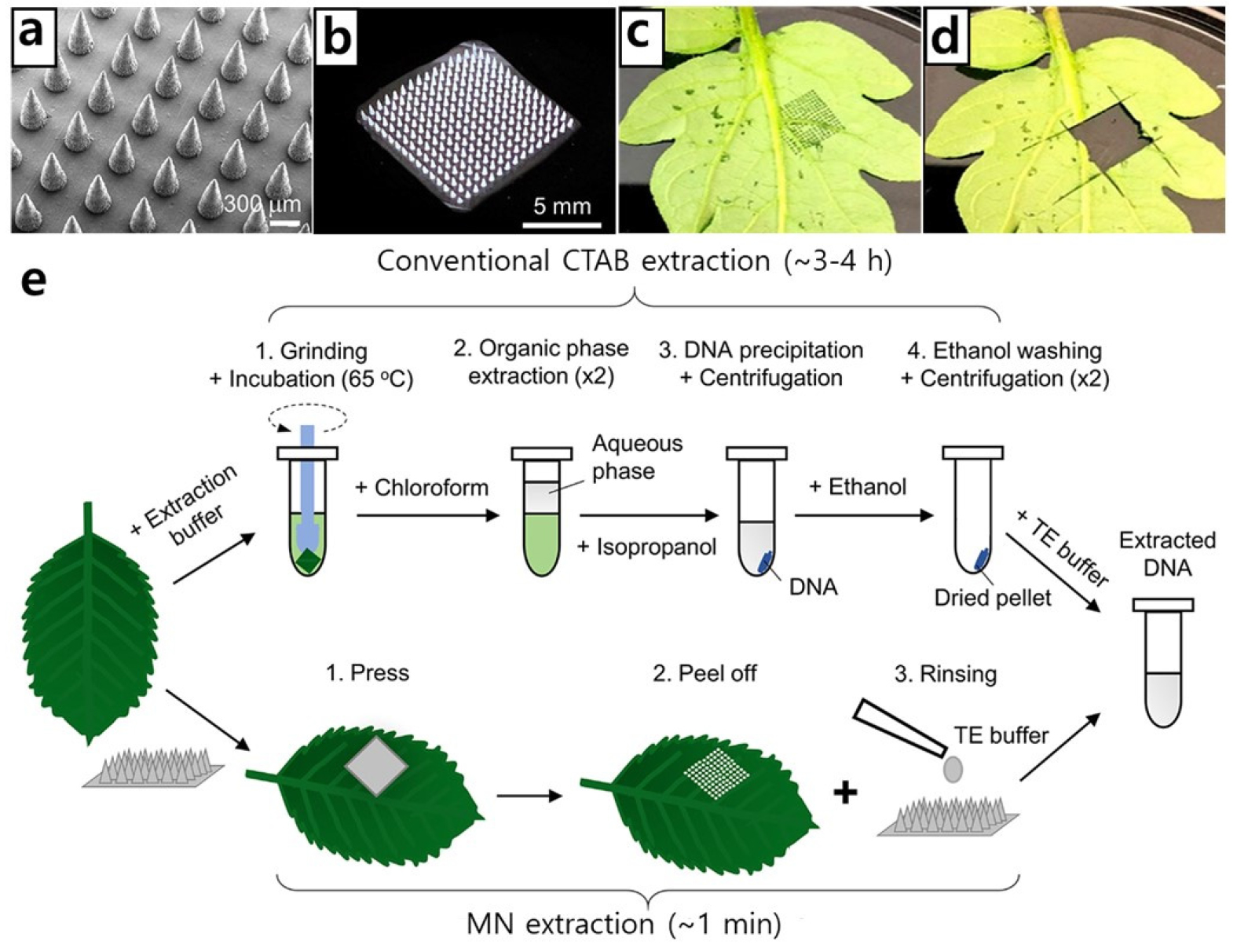
Fig. 2.
a) Scanning electron microscopy (SEM) image of MN patch, b) digital image of the MN patch, c) tomato leaf after the puncture by MN patch, d) tomato leaf after cutting off the area for CTAB extraction, e) schematic representation of conventional CTAB and MN extraction of DNA from leaf (Paul et al., 2019).
Smartphone-based diagnostic tools are now a desirable choice for performing isothermal nucleic acid amplified (NAA) assays in point-of-care sites due to the low cost and widespread availability of smartphones. This has led to the integration of MN extraction with loop-mediated isothermal amplification (LAMP) cassettes and 3D-printed smartphone-based fluorescent reader devices for ‘sample-to-answer’ analysis of plant pathogens (Paul et al., 2021). In this approach, the MN patch utilizes the retraction process to quickly separate the nucleic acids from the plant leaves in just over one minute. The harvested nucleic acids are then fed into an assay cassette to perform an isothermal amplification test, such as LAMP, which contains pre-loaded assay chemicals. Fluorescent images of the LAMP cassette are captured by a smartphone reader device, which detects nucleic acids with a sensitivity of 1 pg. This platform offers a portable approach to plant disease management, enabling rapid and precise detection of pathogens within 30 min without the need for a laboratory. Moreover, this technology can detect tomato wilt virus as early as 5 days post-inoculation in asymptomatic plants. Despite the platform’s promise of enabling rapid on-site pathogen detection, further validation studies across various plant species, environmental conditions, and pathogen concentrations are necessary to ensure its reliability and accuracy.
Detection of signaling molecules
Advancements in precision agriculture rely heavily on prompt access to plant physiological information and real-time monitoring of changes in plant glucose content. To detect glucose in plants minimally invasively, an electrochemical detection device that combines MN sensor and 3D printing technology has been developed (Chen et al., 2024). This device features a 3D-printed polymer hollow MN combined with a platinum wire. The MN incorporates layers of gold nanoparticles, Nafion, glucose oxidase (GOx), and polyurethane (PU) to enhance its sensing abilities. The use of 3D printing technology ensures a slender tip diameter of just 300 µm, reducing plant injury during the recognition process. The MN sensor demonstrates excellent recognition performance, with a limit of detection (LOD) as low as 33.3 µM and detection sensitivity of 17 nA/µM·cm2, enabling the detection of glucose levels in a wide concentration range from 100 µM to 100 mM. When the plant sample interacts with the biosensor, glucose molecules in the plant tissue are catalyzed by the GOx enzyme, producing gluconic acid and hydrogen peroxide (H2O2). This enzymatic reaction produces a biological signal, which is converted into an electric current by the platinum wire. The biosensor detects this current, enabling precise measurement of glucose levels in plant samples. However, it is vital to note that the biosensor’s selectivity for glucose may pose issues when assessing samples containing a various chemical, potentially leading to interference. Further refinement of the biosensor’s selectivity is necessary to ensure accurate measurements in complex plant tissue environments.
H2O2 is a key signaling chemical in plants, playing a vital role in indicating physiological conditions and stress reactions. Monitoring H2O2 concentrations is therefore plants essential for understanding plant health. In a recent study, a PEG-crosslinked poly (methyl vinyl ether-alt-maleic acid (PMVE/MA) hydrogel MN patch was developed for in-field sampling, demonstrated rapid H2O2 sensing (Fig. 3) (Wu et al., 2024). These MNs were developed using the micro molding method, resulting in hydrogel MNs with a round shape and a diameter of 17.5 mm, which facilitated quick extraction of sap from plant leaves. For H2O2 detection, optical detection technology was employed using a portable colorimeter. The colorimeter measures the absorbance of light by the extracted sap samples, allowing for the quantification of H2O2 levels based on observed color changes. When the sap containing H2O2 reacted with a specific reagent in the colorimeter, a color change occurred, corresponding to the concentration of H2O2. This was then compared to the known standard sample. This method provides a rapid, non-invasive, and accurate means of assessing H2O2 levels in plants, offering a practical and efficient way to monitor plant stress responses in real time. The ability to quickly and accurately measure H2O2 levels in the field enhances our capability to manage plant health and stress, ensuring timely interventions and better crop management.
In a study led by Parrilla and group, hollow MN arrays (HMNs) were fabricated using a high resolution 3D printer based on stereolithography (Parrilla et al., 2024). These designed MNs exhibited a tip diameter of 25.9 ± 3.7 µm and a side hole diameter on the MN of 228.2 ± 18.6 µm, allowing for the extraction of > 10 µL apoplast fluid. The HMNs were integrated with graphite based screen-printed electrodes (SPEs) modified with specific materials such as Prussian blue (PB), PB with Gox, and polyaniline (PANI) to detect H2O2, glucose, and pH, respectively. These sensors are capable of gently penetrating plant tissues to access apoplast fluid, enabling the collection of samples for precise analysis. Within the sensing system, a paper-based electrochemical cell assists in allowing smaller molecules to reach electrodes while trapping larger interfering molecules that could disrupt electrochemical reactions. PB, for instance, facilitates the reduction of H2O2, generating a measurable current directly proportional to the H2O2 concentration. Similarly, GOx catalyzes the oxidation of glucose, with the resulting product detected electrochemically. The pH sensor measures the proton concentration, correlating it with the pH of the sample. However, the broad applicability of the system to detect a wide range of biomarkers may be influenced by the sensitivity and selectivity of the integrated electrochemical sensors. Variations in these factors could potentially impact the accuracy of the measurements obtained. Further refinement and validation of the sensor technologies are necessary to ensure robust performance across diverse plant samples and environmental conditions.
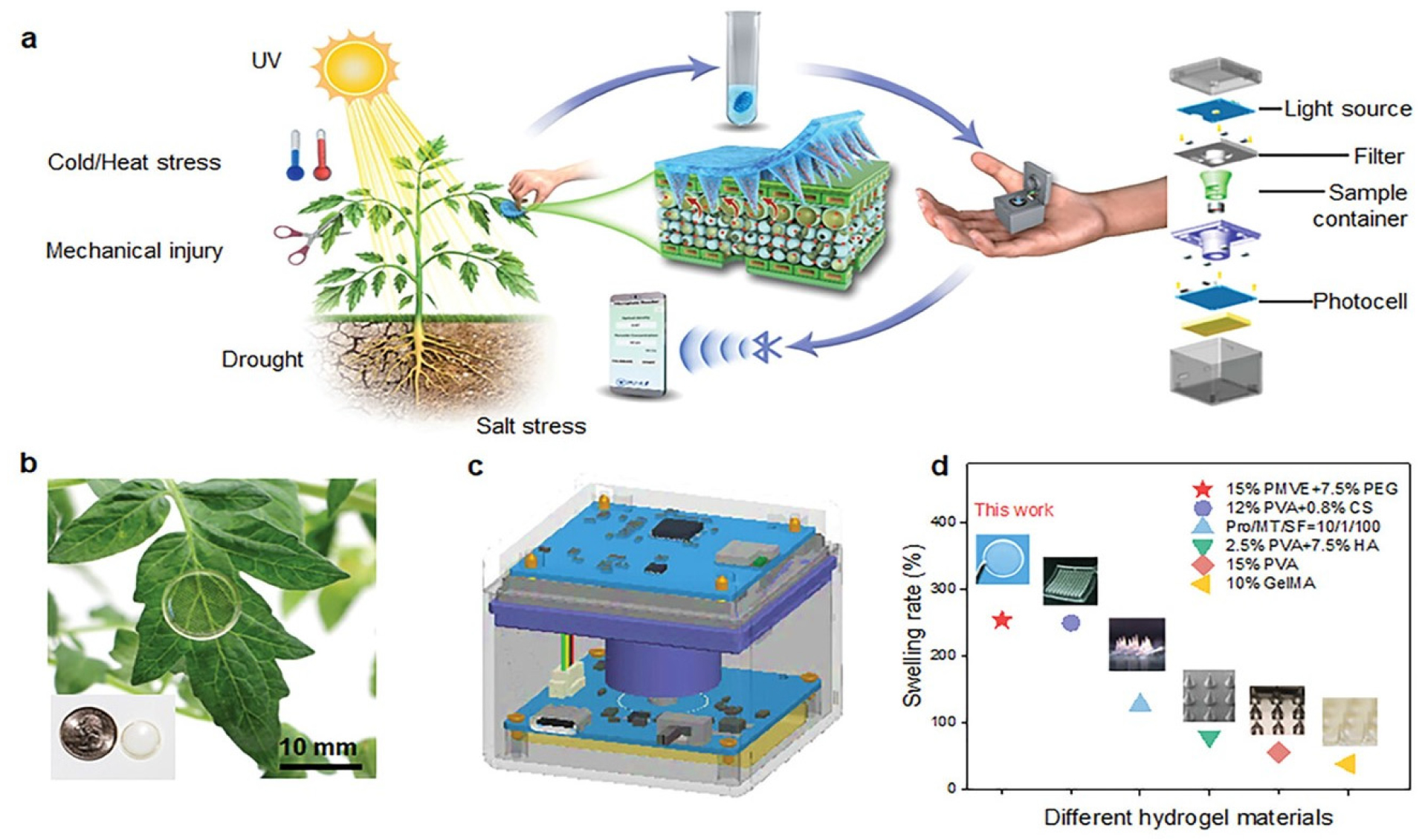
Fig. 3.
a) PEG-crosslinked PMVE/MA Hydrogel MN patch, b) photography of MN patches employed on a plant leaf, c) structure of colorimeter, d) swelling rates of different MN materials (Wu et al., 2024).
Detecting metabolites
The analysis of abscisic acid (ABA) in trace amounts within plants is crucial due to its significant role as a natural growth that inhibits plant growth (Kishor et al. 2022). To facilitate the monitoring of this metabolite, a MN array sensor has been developed, featuring microelectrodes made from Au@SnO2-vertical graphene (VG) on a tantalum (Ta) substrate (Wang et al., 2021). Herein, VG film acts as an electrocatalyst, while Au@SnO2 nanoparticles serve as specific active sites for detecting ABA. The Au@SnO2-VG layers were directly grown on Ta wires using direct current (DC) arc plasma jet chemical vapor deposition (CVD), enabling quantitative detection of ABA through direct electrocatalytic oxidation. Upon exposure to ABA in plants, the Au@SnO2 nanoparticles on VG nanosheets catalyze the electrocatalytic oxidation process, leveraging the numerous catalytic active sites provided by the nanoparticles combined with the excellent conductivity of VG nanosheets. The sensor operates effectively within a pH range from 4 to 7, offering a response concentration range spanning from 0.012 (or 0.024) to 495.2 µM, and a detection limit ranging between 0.002 and 0.005 µM. Despite demonstrating high selectivity, repeatability, reproducibility, and long-term stability, potential issues remain regarding cross-reactivity with other compounds present in plant tissues that could potentially interfere with accurate ABA detection. Addressing these concerns through further validation studies under diverse environmental conditions and with various plant species will be essential to ensure the sensor’s reliability and applicability in practical agricultural settings.
Plants adjust to environmental stresses primarily through phytohormone facilitated regulation of oxidative stress. While there is evidence correlating stressors with phytohormone levels, understanding the mechanism of interaction among phytohormones is hindered by the lack of in situ sensing technology. Addressing this research gap, a bioagent-free, stem-mounted MN based sensor with an implanted pH sensor on the same chip was constructed to monitor salicylic acid (SA) and pH levels from the plant sap (Hossain and Tabassum, 2022). The sensor’s working electrode was modified using a copper metal-organic framework, while the pH sensor was coated with a pH-selective material, PANI. Differential pulse voltammetry (DPV) was employed to measure SA levels, showing an increase in current corresponding to the SA peak as the SA concentrations in the plant sap increased due to the SA oxidation by the Cu-MOF coating. The sensor displayed a robust linear response with a high sensitivity and a low detection limit, capable of detecting SA levels as low as 37 µM. Notably, the sensor accurately captured dynamic variations in SA readings within the stems of both water-stressed and unstressed plants, providing insights into plant responses to different stressors. In addition, advancements in multiplexed recognition of other defense-related phytohormones are crucial to monitoring real-time hormonal variations comprehensively.
Monitoring physiological conditions
The integration of physiological knowledge into agricultural practices is pivotal for enhancing the efficiency, sustainability, and productivity of farming systems. Monitoring the xylem sap flow plays a crucial role in understanding plant physiology, as it regulates plant response to environmental changes such as soil water content, humidity, and sunlight (O’brien et al., 2004). To address this need, a MN sensor was fabricated, involving the deposition of titanium and gold layers using e-gun evaporation as an essential component (Baek et al., 2018). The sensors were further enhanced with a top insulation layer of silicon nitride deposited via plasma-enhanced chemical vapor deposition, ensuring protection and functionality. The MN structure essential for accurate measurement of sap flow rate was achieved through a deep reactive ion etching process on the silicon substrate. When the MN comes into contact with water flow, the integrated heating element in the sensing system activates, generating localized heat around the MN. measurement circuits capture temperature variations over specific time intervals, converting these fluctuations into data that quantify the rate of water transport in the plant stem. However, the fabrication process involving complex techniques like e-gun evaporation and plasma-enhanced chemical vapor deposition may present challenges in scalability and cost-effectiveness for widespread implementation. Efforts to streamline manufacturing processes and explore alternative fabrication methods could potentially mitigate these challenges, making the technology more accessible for broader adoption in agriculture.
Understanding plant structural changes that reflect physiological conditions, such as differences in ionic content, membrane permeability, and viscosity, provides insights into plant nutrition, stress responses, and overall health. Monitoring these changes is essential for enhancing plant resilience and productivity in agriculture. A recent study by Bukhamsin et al. (Bukhamsin et al., 2021) introduced impedimetric sensors employing MNs, designed to pierce the waxy exterior of plants and generate sensitive impedance spectra in open-air conditions (Fig. 4). The MNs were fabricated using the micro-molding method and integrated into the sensor system with silver paste coated copper wire electrodes. These MNs penetrate the plant’s waxy external layer, establishing direct electrical contact with internal tissues while minimizing damage to the plant. Importantly, the sensor’s performance was validated through experiments on Arabidopsis thaliana, demonstrating its ability to detect impedance changes influenced by environmental factors such as light exposure and hydration levels. This highlights its potential for long term deployment in precision farming, where continuous monitoring of plant health and responses to environmental stimuli is critical.

Fig. 4.
a) Schematic representation showing the impedance sensor employed to plant leaf, b) photography of polyimide MNs after demolding, c) SU-8 MNs after demolding, d&e) SEM images of the polyimide MNs at different magnifications (Bukhamsin et al., 2021).
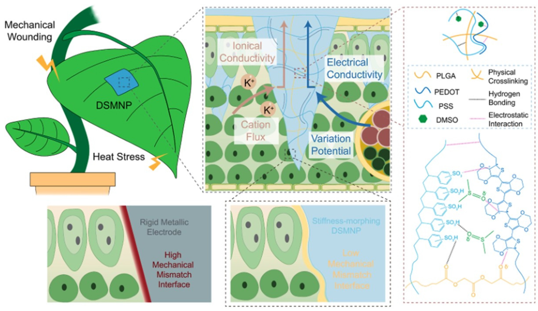
In another study addressing the limitations of traditional molecular and cellular biology techniques, Kong et. al. introduced a dual-conductive and stiffness-morphing MN patch (DSMNP) designed for continuous monitoring of electrophysiological signals and ion fluctuations in plants (Fig. 5) (Kong et al., 2023). The MN patch was fabricated using a combination of photolithography, electrodeposition, and micro-molding techniques with PDMS serving as a flexible substrate. These MNs can penetrate plant tissues to establish intimate contact with the vascular system without causing significant damage or stress. The primary sensing mechanism relies on electrochemical methods; once inserted into the plant, the MN detects the electrical signals indicative of water content and ionic concentration. This detection leverages the MN conductive properties to capture bioelectrical signals associated with the plant’s water status. Collected electrical signals are transmitted to an external data acquisition system for processing and analysis enabling continuous monitoring of the plant’s water stress levels. This innovative sensing approach provides precise and non-invasive assessment of plant water stress, making a significant advancement in agricultural technology.
In a study aimed at examining the bioimpedance of Barley leaves under controlled conditions, Bukhamsin et al. developed flexible and lightweight MN plant electrodes (Bukhamsin et al., 2020). These biosensors were produced using a PDMS imprinting method with a 3D printed negative mold, demonstrating versatility in achieving MN heights up to 500 µm while maintaining high fidelity to the desired MN geometry. The objective was to enable rapid, real-time, and non-destructive monitoring of crops, potentially enhancing field productivity and reducing resource consumption in agriculture. Comparative performance assessments between the MN electrodes and traditional planar electrodes were conducted to measure the bioimpedance of plant tissues. The fabrication process involved PDMS utilization, laser lithography for precise mold creation, and metal coating to enhance MN mechanical strength. Measurements revealed diurnal variations in barley bioimpedance at low frequencies, likely influenced by light cycles. Future work should focus on using this technique to catalog the responses of barley and other commercially significant crops to various abiotic and biotic stress factors. Future research directions could explore applying this technique to systematically document how barley and other economically significant crops respond to diverse biotic and abiotic stressors.
Jeon et al. introduced a MN structure integrated with electrodes to precisely measure impedance, aiming to understand plant responses to nutrient solutions (Jeon et al., 2017). Fabricated through silicon etching and micro-patterning, the MN featured a height of 500 µm and a width of 200 µm. In experiments conducted on tomato stems, real-time impedance measurements were converted into electrical conductivity (EC), enabling continuous monitoring of how plants react to external variables like temperature fluctuations and nutrient intake. This real-time EC measurement of tomato sap provides insights crucial for assessing plant health and optimizing nutrient solution salinity. By constructing growth models based on these EC measurements, researchers can better comprehend how plants respond to environmental conditions. Ultimately, this contributes to refining agricultural practices and enhancing crop cultivation outcomes. Future studies could further refine the sensor’s accuracy and explore its application across various plant species and environmental settings to broaden its utility in precision agriculture. Table 2 summarizes the recent MN sensor technology, parameters, and applications in precision agriculture.
Table 2.
Various MN biosensors for plant health monitoring.
| MN type | Design and structure | Target | Purpose | Result | Ref. |
| PVA based MN |
Conical cavity structure with a height of 800 µm, a base radius of 150 µm, and a tip radius of 5 µm | Tomato leaf |
Late blight disease caused by P. infestans |
Allowed DNA extraction within 1 min and yielded a detection rate of 100% | (Paul et al., 2019) |
| PVA based MN |
Conical cavity structure with a height of 800 µm, a base radius of 150 µm, and a tip radius of 5 µm | Tomato leaf |
Late blight disease caused by P. infestans |
Rapid and precise detection of pathogens within 30 min with a sensitivity of 1 pg | (Paul et al., 2021) |
|
3D printed polymer hollow MN |
Hollow structure with a slender tip diameter of 300 µm | Tomato stem | Glucose |
Detection performance with LOD of 33.3 µM and detection sensitivity of 17 nA/µM・cm2 | (Chen et al., 2024) |
|
PEG-crosslinked PMVE/MA hydrogel MN patch |
Round shape with a diameter of 17.5 mm | Tomato leaf | H2O2 |
Rapid detection of H2O2 content in plant leaves with minimal plant damage | (Wu et al., 2024) |
|
3D printed hollow MN arrays |
Hollow structure with a tip diameter of 25.9 ± 3.7 µm and a side hole diameter on the MN of 228.2 ± 18.6 µm | P. peperomioide leaf |
H2O2, glucose, and pH |
Efficient passive fluid extraction with rapid on-site plant health monitoring | (Parrilla et al., 2024) |
| Au@SnO2-VG MN |
Consists of a polytetrafluoroethylene fixing clip with the size of 15 mm × 7 mm × 7 mm | Cucumber | Abscisic acid |
Operates effectively across a pH range from 4 to 7 with a detection limit ranging between 0.002 to 0.005 µM | (Wang et al., 2021) |
| Resin based MN |
Pyramidal shaped with a square base of 800 µm and a height of 2000 µm | Cabbage plant | Salicylic acid |
Strong linear response with a high sensitivity and a LOD of 37 µM | (Hossain and Tabassum, 2022) |
| Silicon MN |
Sensing probe 5 mm long, 500 µm wide, and 300 µm thick, two auxiliary needles, contact pads, and multiple heaters | Tomato stem | Water transport |
Effectively measured the water flow speed using a MN thermal probe | (Baek et al., 2018) |
| Polymeric MNs |
Conical in shape with a radius of 70 µm and a height of 350 µm | A. thaliana |
Electrical impedance |
Obtained sensitive impedance spectra with an average relative noise value of 3.83% and the sensor can be used for 20 replication cycles | (Bukhamsin et al., 2021) |
|
Poly(3,4-ethylene dioxythiophene), polystyrene sulfonate, poly(lactic-co-gly colic acid) based MNs |
Pyramidic MN with the tip size of ~10 µm, length of 500 µm, base dimension of 250 µm, and inter-MN spacing of 400 µm | E. aureum |
Electrophysiological signals and ion fluctuations |
Conductive property of MN facilitates the measurement of electrical signals | (Kong et al., 2023) |
Conclusion and future prospective
MN technology presents a transformative approach to enhancing plant health management and agricultural diagnostics. Traditional monitoring methods often require destructive sampling, which is time-consuming and impractical for real-time field applications. In contrast, MN devices enable noninvasive monitoring and efficient sample extraction, drastically reducing analysis time from hours to minutes. These devices offer several advantages, including rapid on-site diagnostics of plant health, allowing farmers to swiftly implement treatment strategies without the delays associated with laboratory testing. By minimizing sample usage and maintaining sensitivity, MN based sensors offer cost-effective solutions that can be easily deployed even by non-specialists in the field. Despite significant advancements, challenges remain in scaling up manufacturing to ensure cost-effectiveness and widespread commercial availability of MN devices. Additionally, the stability of microneedles (MNs) over time when exposed to environmental conditions is a concern, as it may lead to inaccurate sensor readings. The presence of organic matter in the application area can also interfere with MN sensor data. Moreover, the environmental impact of disposing of used or damaged MNs, especially those containing hazardous materials, needs careful consideration. The potential toxicity of materials used in these sensors to plants, soil organisms, and the broader ecosystem must also be thoroughly assessed. Large size MNs can cause more significant damage to plant tissues during insertion. This can weaken the plant and potentially introduce pathways for infections or stress, altering its physiological state temporarily or even causing lasting damage if not carefully managed. Addressing these challenges is crucial for realizing the full potential of this technology in precision agriculture. Despite significant advancements in the field, MN sensors have yet to be commercialized. The primary hurdle is the challenge of cost-effective, large-scale manufacturing. Despite the difficulties in developing MN devices, this miniaturized and convenient technology holds remarkable potential for enhancing plant and crop disease monitoring, diagnostics, and health management. Looking ahead, the future of smart agricultural biosensing hinges on enhancing sensor sensitivity, reliability, reproducibility, and specificity, while ensuring devices are compact, affordable, and easy to use. By bridging the gap between laboratory precision and field usability, MN devices are expected to play a pivotal role in the next generation of agriculture, fostering smarter and more efficient farming practices.
